
This application of WebRotate 360 began as a project at the Cleveland Museum of Natural History to create online 3D models of insect specimens, enabling easy examination for scientific research. These specimens are normally stored in museum collections and are available to researchers who can afford the time and expense to visit the collection, or they can be shipped to the researcher at the risk of loss or damage to the fragile specimens in transit.
Online specimen models would be safely available anytime, anywhere, and to anyone for free.
3D models of real objects are usually produced using 360-degree photogrammetry or by 3D scanning. The latter technique is very fast but a micro 3D scanner is expensive, and has limitations in the resolution of tiny details, as well as difficulty with transparent or shiny surfaces. Producing a 3D model from photogrammetry typically involves processing the camera images through meshing and modeling software to create a rotatable screen object. In our experience this approach results in a loss of high-resolution photographic details that are important for scientific analysis of specimens.
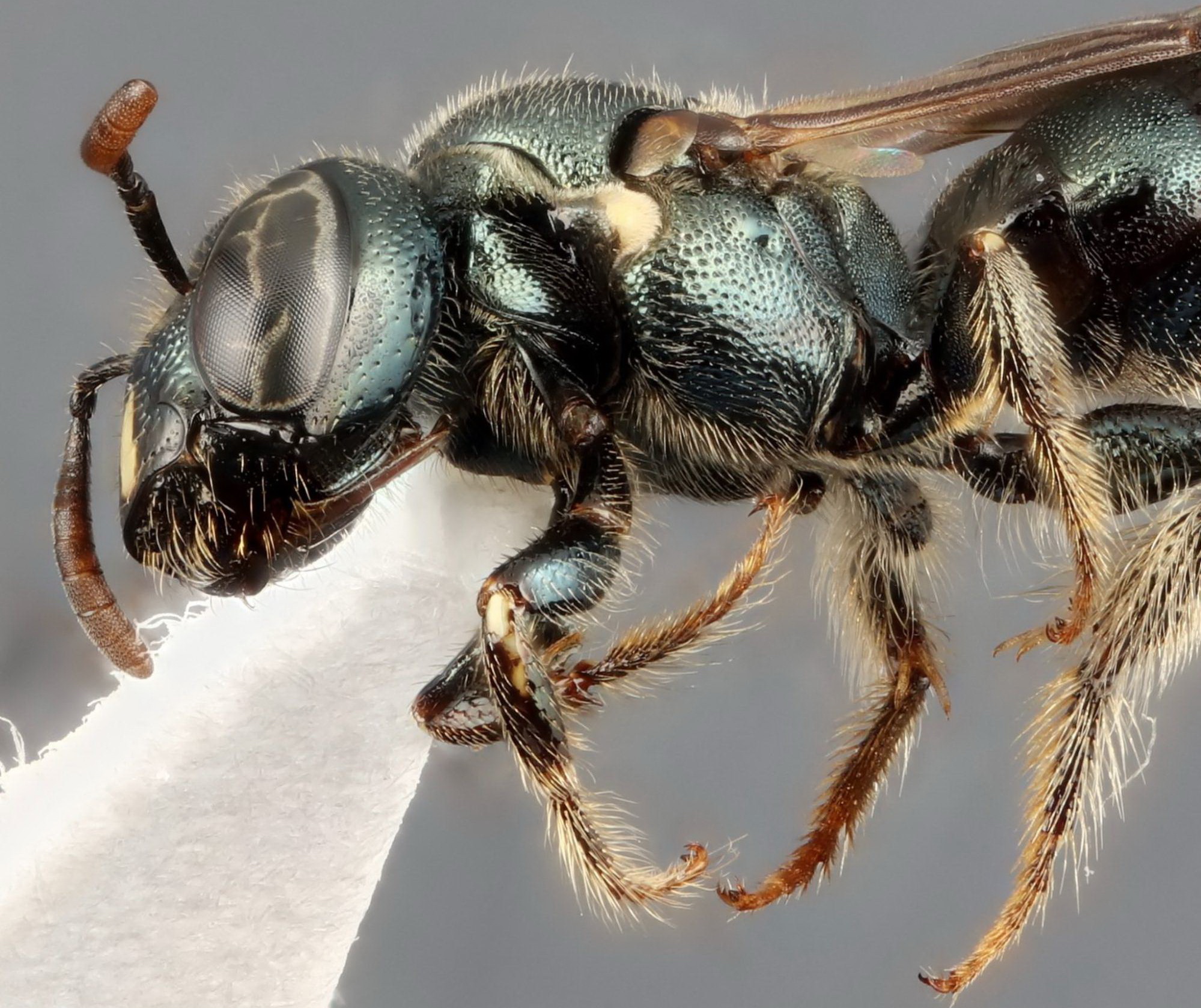 Zoomed image provides microscopic detail not seen in 3D models
This is where WebRotate 360 excels for our application. We are able to create screen models of insects using multiple 2D macro images that retain all of the high-resolution details and simulate looking at the actual specimen under a microscope – a "virtual specimen".
The process we use is time-consuming, but almost entirely automated. We shoot between 182 and 254 views of each specimen as it is rotated and tilted at specified angles. Because the insects are so small (typically 3 - 50 mm), each one of the views requires focus-stacking 10-40 macro images with extremely shallow depth of field. It can take up to 10,000 photos and 4 hours to shoot one specimen, although most are half that.
We’re using a Canon 5DS R camera for maximum sensor resolution (50MP) with macro lenses attached to a motorized focus-stacking rail, with the specimen mounted (using its existing mounting pin) on a 360° motorized turntable. This in turn is tilted on a motorized arm for 5-7 rows of images. The motors are programmed and controlled with a StackShot 3X from Cognisys.
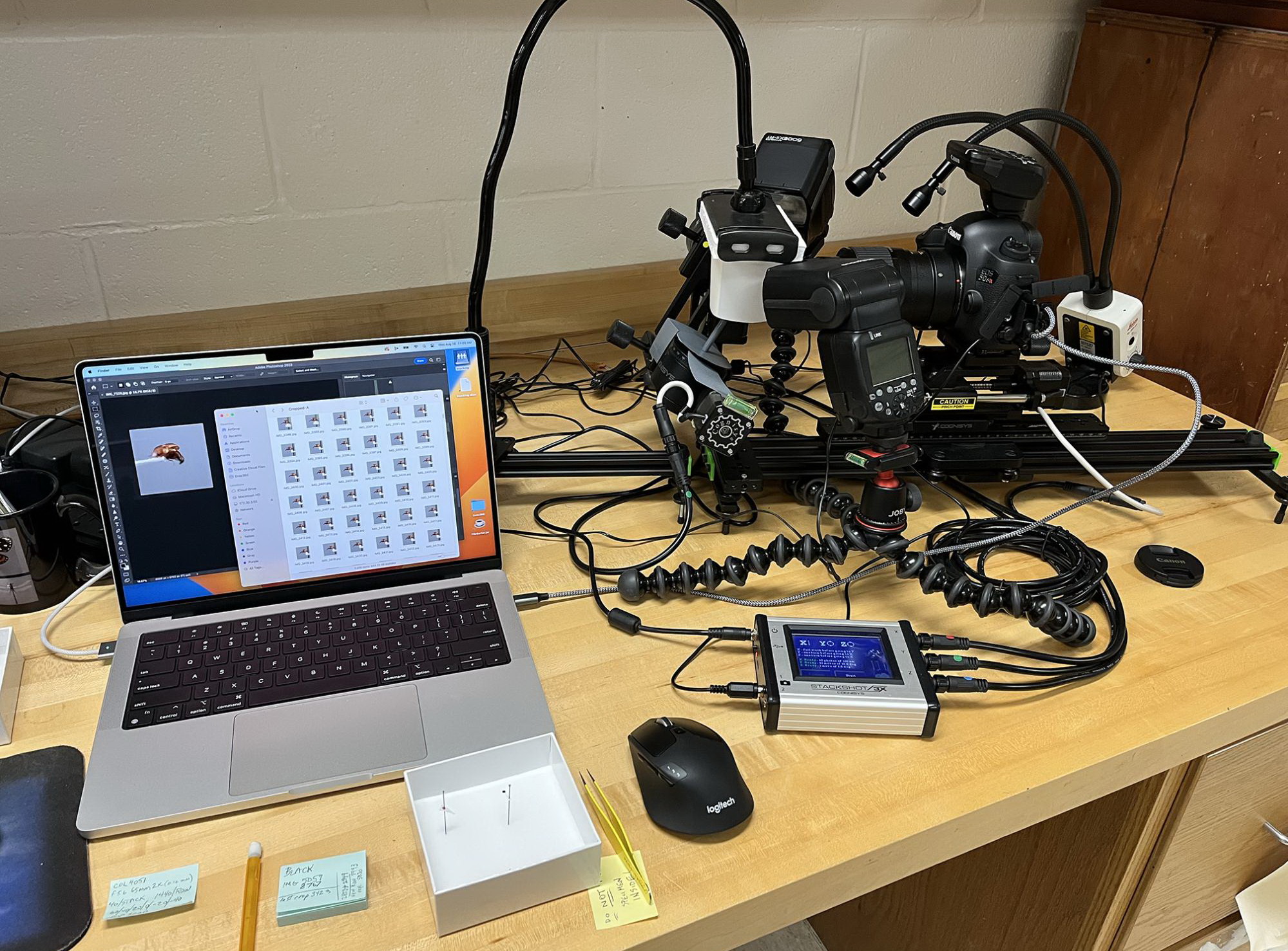 Full rig with laptop for museum travel
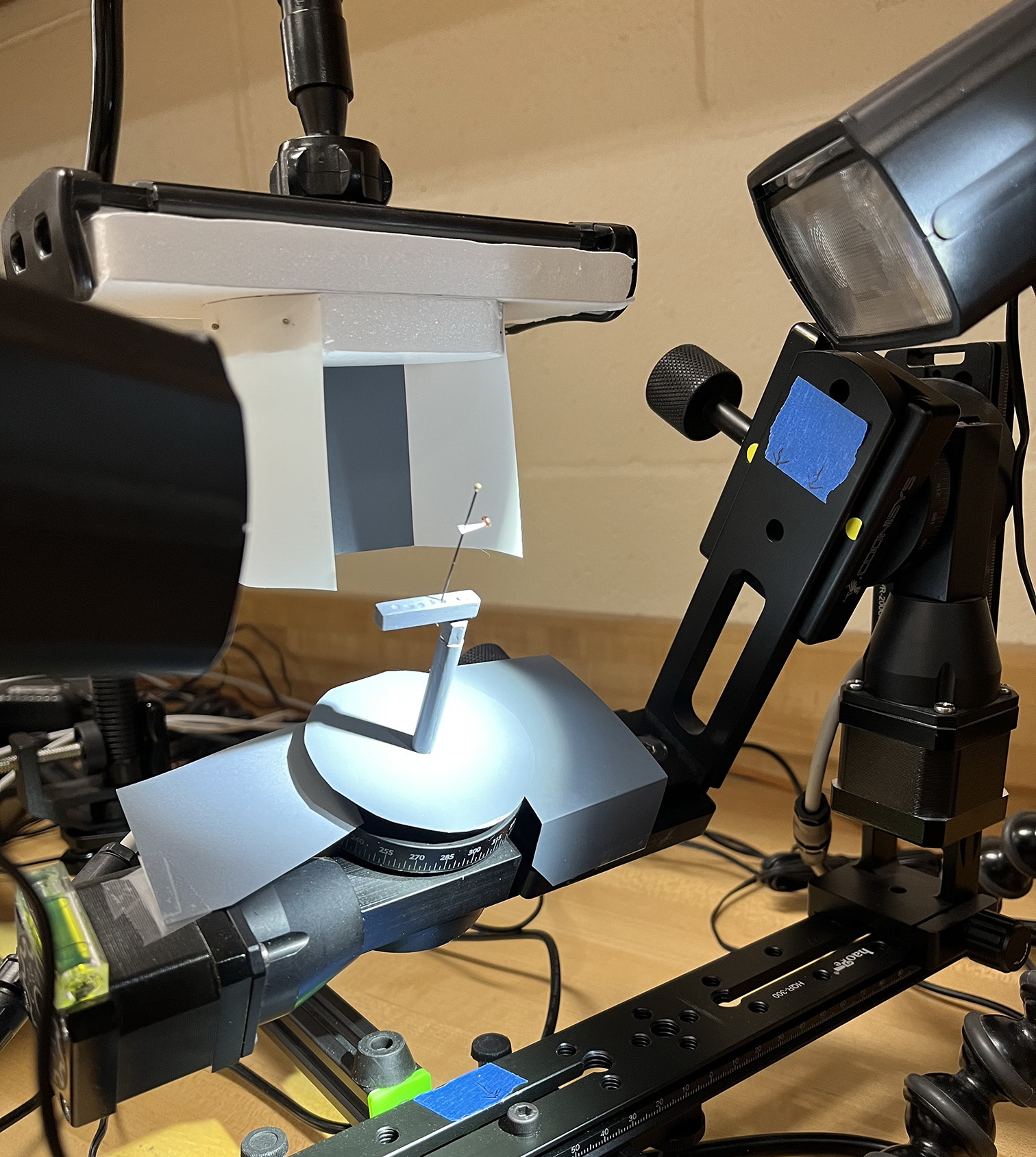 Tilted Z-axis with view of flash diffuser drawn back on its own rail to view specimen. The Z-axis is tilted in 20-30 degree increments, depending on the specimen. Also visible is a special mount to accommodate insects with off-center pins (such as the tiny "point-mounted" beetle shown here)
The 50-megapixel images are then batch-processed over several more hours (or concurrently) with Zerene stacking software and Photoshop to create the cropped views that are imported into WebRotate 360 – retaining 3000-4000 pixel resolution for online magnification.
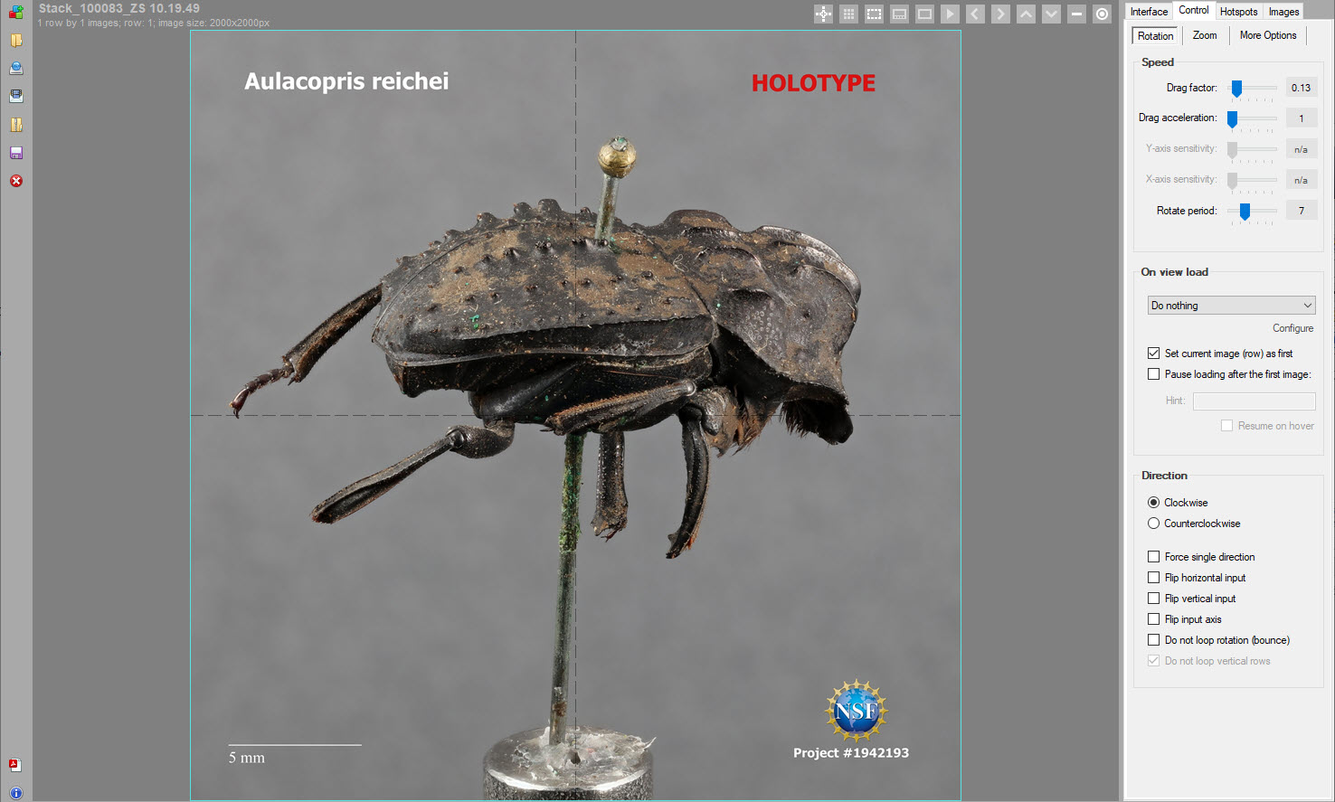 360-degree images are imported into WebRotate 360 software for 360 view configuration and publishing
The final web model (example) is then created in a matter of minutes, after adding canvas text, logo, and hotspots. Top and bottom (dorsal and ventral) views of the specimen are created, respectively, from a single focus-stacked image which is rotated in 10° increments in Photoshop to create the 36 views that fill the first and last rows of the WebRotate 360 import.
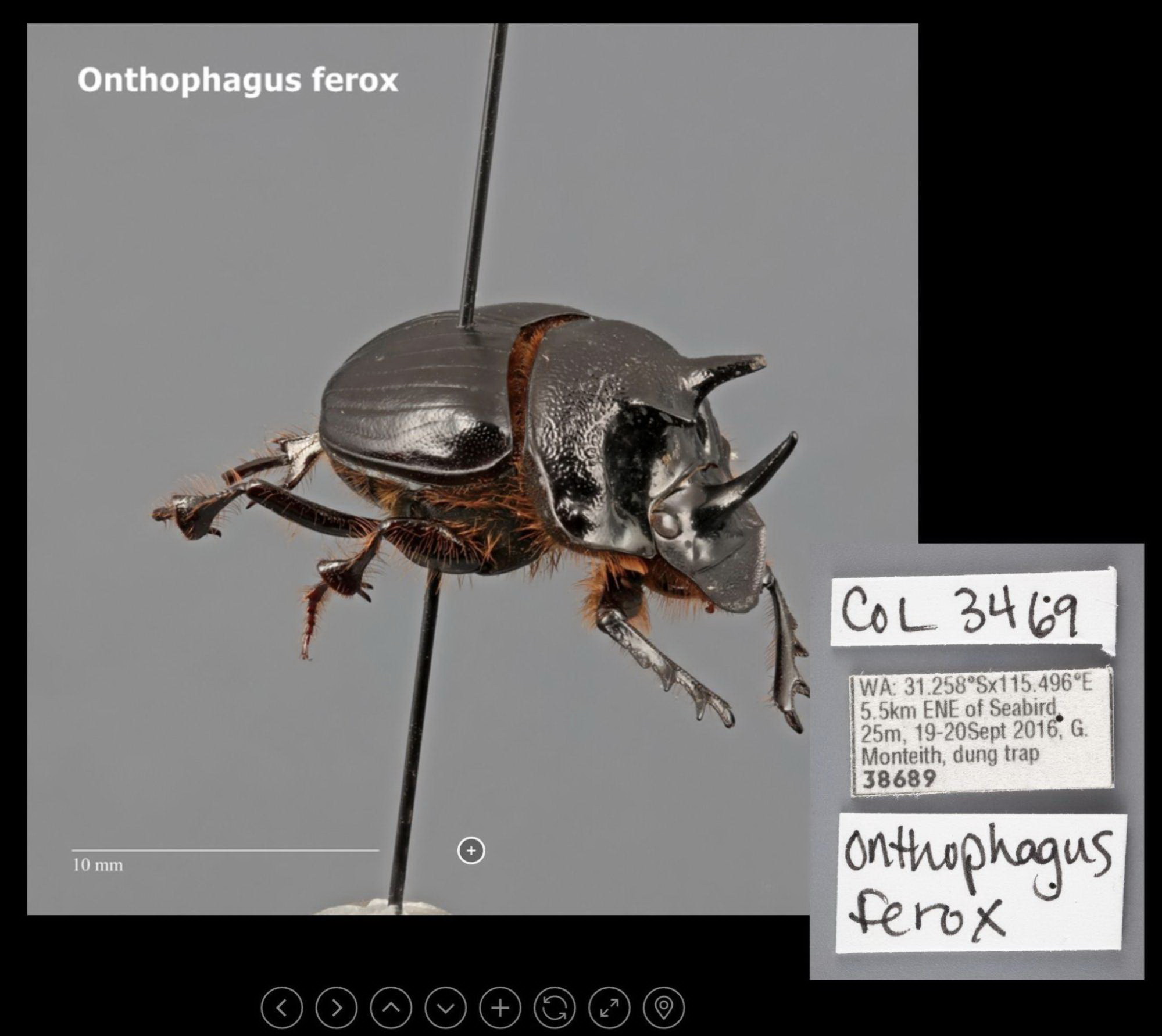 Hotspot button shows popup with insect’s pin labels
The resulting web site interface provides easy controls for manipulating the virtual specimen and zooming in for full-resolution details. A hotspot button provides an image of the identifying labels that are kept on the insect’s pin in the museum’s collection drawer.
We have currently imaged over 150 specimens, which can be viewed online at Ento360.